

Tracheobronchomalacia (TBM) is a condition characterized by cartilage weakening of the tracheal or bronchial wall. This condition causes tracheal and bronchial collapse resulting in airflow obstruction.
The TBM is treated by positioning an internal stent to support the weakened wall and prevent collapse.
In this scenario a tracheobronchial model has been reproduced with the purpose of being used for physicians training in two different tasks:
1) Endoscopic examination
2) Internal stent application
The simulator was created from CT images of a patient affected by tracheomalacia. The figure shows the virtual 3D reconstruction of the anatomical model (1.a) and the physical replica obtained with the 3D printer (1.b). In addition to the trachea, the simulator also includes parts of surrounding anatomical regions such as the pulmonary artery, aortic arch and esophagus.
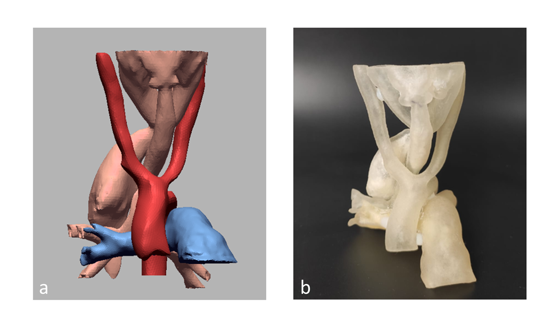
Figure 1: Anatomical model 3D reconstruction. a) Virtual reconstruction b) Physical replica