Atrial septal defect (ASD) is a congenital heart defect in which the atrial septum that normally separates the right atrium from the left atrium of the heart is open, with a subsequent blood flow. Thus, the oxygen-rich blood can flow directly in the right side of the heart to mix with the oxygen-poor blood or the opposite. This can lead to lower-than-normal oxygen levels in the arterial blood.
The ASD closure can be obtained in percutaneous way, by means of a specific occluder (ASO – AMPLATZER Septal Occluder). The occluder, which assumes the shape of a little umbrella when opened, is introduced in the atrial zone passing through a vein (generally, the femoral vein).
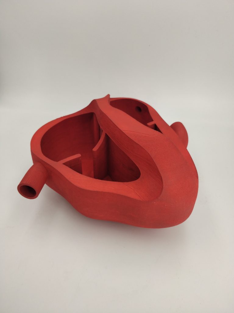
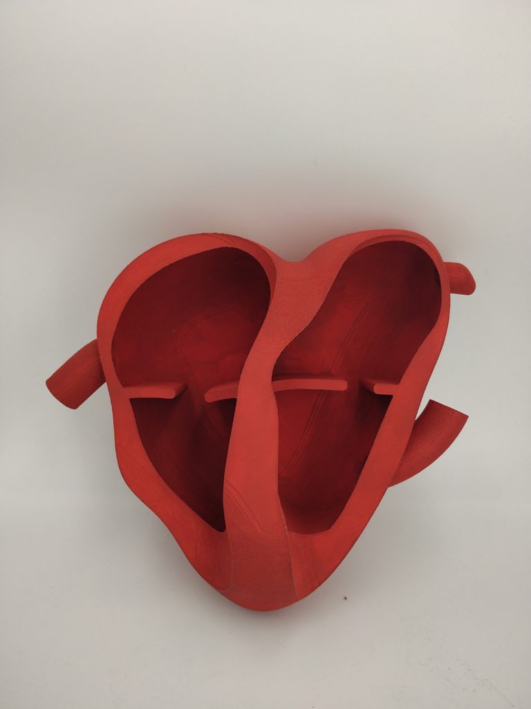
The 3D informative model helps the cardiologist in explaining the procedure to the patient, by means of simulation.
Modeling
The 3D model is composed by 2 components: 1. 3D simplified heart model and 2. 3D simplified removable septum model. The latter – in the area that is subject to the intervetion – is made of silicon to reproduce the high flexibility of the soft tissue and make the defect occlusal realistic. The septum model is removable to allow a fast and easy replacement in case of excessive usage.
The heart modeling is intentionally simplified to give the patient an optimal view of the area of intervention.